Call Today! 646-854-5736
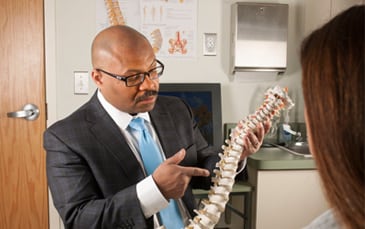
Dr. Senatus is a Highly Awarded, 5 Star reviewed Board-Certified Neurosurgeon Specializing in Minimally Invasive Spine Surgery.
Located on the Upper East Side of Manhattan.
5-Star Rated Neurosurgeon

Why Choose Dr. Senatus?
Dr. Patrick Senatus is a Board Certified and Fellowship Trained Neurosurgeon with an expertise in Minimally Invasive Spinal Surgery. He also serves as Assistant Clinical Professor of Neurological Surgery at Yale School of Medicine.
Dr. Patrick Senatus attended Harvard where he was recognized as a Harvard College and John Harvard Scholar and graduated Magna Cum Laude with a Bachelor of Art in Biochemical Sciences. As a Medical Scientist Training Program Scholar, he completed his Doctorate of Medicine at Harvard Medical School and his Doctorate of Philosophy in Neurobiology at the Harvard Graduate School of Arts and Sciences.
In 2006, Dr. Senatus completed a Neurosurgery Residency at Columbia Presbyterian Medical Center where he was nominated for the Arnold P. Gold and Physician of the Year Awards. Subsequently, he completed a Fellowship at the Cleveland Clinic in Functional and Restorative Neurosurgery of the Spine and Brain.
Click Below To See The Conditions We Treat:
- Chronic Pain
- Lumbar Herniated Disc
- Cervical Herniated Disc
- Sciatica
- Spinal Stenosis
- Cervical Radiculopathy
- Cervical Myelopathy
- Scoliosis
- Ankylosing Spondylitis
- Spondylolisthesis
- Compression Fracture of the Spine
There are many different conditions that cause or contribute to low and lower back pain. Some of these spinal conditions are listed below.
- Bulging or herniated disc. A disc may bulge outward. A herniated disc occurs when the soft interior matter escapes through a crack or ruptures through the disc’s protective outer layer. Both disc problems can cause nerve compression, inflammation, and pain.
- Spinal stenosis develops when the spinal canal or a nerve passageway abnormally narrows.
- Spinal arthritis, also called spinal osteoarthritis or spondylosis, is a common degenerative spine problem. It affects the spine’s facet joints and may contribute to the development of bone spurs.
- Spondylolisthesis occurs when a lumbar (low back) vertebral body slips forward over the vertebra below it.
- Vertebral fractures (burst or compression types) are often caused by some type of trauma (eg, fall).
Lumbar Herniated Disc, also known as slipped disc or ruptured disc, is a condition in which the spinal disc begins to protrude into the spinal canal. This protrusion of the disc can irritate the surrounding nerve roots of the spine and cause localized or radiating pain.
What are the Symptoms?
Many patients will show visible disc herniations on an MRI but never experience pain or symptoms and others will experience chronic symptoms that include:
- Lower back pain
- Muscle spasms
- Stiffness
- Sciatica
- Numbness or tingling the legs
Cervical Herniated Disc, also known as slipped disc or ruptured disc, is a condition in which the spinal disc begins to protrude into the spinal canal. This protrusion of the disc can irritate the surrounding nerve roots of the spine and cause localized or radiating pain.
What are the Symptoms?
Many patients will show visible disc herniations on an MRI but never experience pain or symptoms and others will experience chronic symptoms that include:
- Neck pain
- Shoulder pain
- Muscle spasms
- Stiffness
- Radiculopathy
- Numbness or tingling the arms and fingers
Despite being commonly used as a term to describe radiating pain in the buttocks and legs, Sciatica is not an actual diagnosis and is instead a symptom of an underlying spinal condition. The sciatic nerve is the longest continuous nerve in the body and extends from the lumbar spine through the back of each leg. When a spinal disc, piece of bone or other process irritate this nerve it can cause the radiating pain that is referred to as Sciatica.
The most common conditions that cause Sciatica are:
- Lumbar Disc Herniation
- Lumbar Spinal Stenosis
- Degenerative Disc Disease
- Spondylolisthesis
- Spondylosis
- Spinal Arthritis
What are the Symptoms?
Sciatic pain usually occurs at one side of a person’s body and it is very unlikely that if affects both sides of the body. The most common symptoms of sciatica are:
- Burning or radiating pain in the lower back, buttocks, hips or legs
- Weakness in the legs or feet
- Numbness in the legs or feet
Spinal Stenosis is a condition in which the spaces in the spinal cord begin to narrow which consequently increases pressure on the spinal cord and the nerves of the spine. Almost all cases of Spinal Stenosis occur in the lumbar spine (lower back). Spinal Stenosis can occur in the cervical spine, yet is far less likely. Spinal Stenosis causes the impingement of one or more spinal nerve roots, which is the cause of the pain associated with Spinal Stenosis.
What are the Symptoms?
The symptoms of Spinal Stenosis can include:
- Lower back pain
- Muscle spasms
- Stiffness
- Sciatica
- Numbness or tingling the legs
Similar to Sciatica, Cervical Radiculopathy is a term used to describe radiating pain that originates in the cervical spine (neck) and radiates into the extremities, but is not an actual diagnosis. When a spinal disc, piece of bone or other process irritates a nerve in the cervical spine it can cause the radiating pain that is referred to as Radiculopathy.
What are the Causes?
Cervical Radiculopathy is always caused by an underlying cervical spine condition and the most common conditions that result in Cervical Radiculopathy are:
- Cervical Disc Herniation
- Degenerative Disc Disease
- Spondylolisthesis
- Spondylosis
- Scoliosis
- Spinal Arthritis
Cervical Myelopathy is a spinal cord disease or dysfunction resulting from abnormal pressure being placed on the spinal cord. This condition can have several causes and the most common is Cervical Disc Degeneration. As we age, the intervertebral discs begin to lose height, which can cause the vertebrae above and below to rub together. As the vertebrae rub against one another bone spurs (osteophytes) can begin to form and impinge the spinal cord and narrow the spinal canal.
Rheumatoid arthritis has also been linked to Cervical Myelopathy. With RA, the body will begin to attack its own tissue, which causes the tissue to swell and compress the spinal cord.
What are the Symptoms?
- Stiffness or pain in the neck
- Pain that radiates into the shoulders, arms, hands or fingers
- Difficulty with fine motor skills such as writing or grasping objects
- Pain that is worsened when moving the head forward
- Problems with coordination
- Difficulty walking
Scoliosis is a condition in which the straight vertical line of the spine develops a curvature toward either side. From the side view, the spine shows a slight roundness in the upper back, and a degree of inward curvature in the lower back. The spine of a person with scoliosis appears to be curved when viewed from the back or front.
What are the Symptoms?
The most evident symptom of scoliosis is the unusual curvature of the spine. The curve may not be very noticeable if it’s mild. However, the affected person may have an uneven waist, or a one shoulder being higher than the other.
Some other symptoms include:
- Chest pain
- Shortness of breath
- Rib pain
- Back pain
- Abdominal pain
- Muscle Spasms
- Neck pain
This chronic (long term) condition is a type of arthritis that affects the spine. As a result of increased bone spurs (osteophytes) in the spine, the vertebrae may fuse together or grow, causing a rigid spine. The symptoms of Ankylosing Spondylitis may develop over a period of many months or even years.
What are the Symptoms?
The initial symptoms and signs of Ankylosing Spondylitis include stiffness and pain in the hips and lower back, particularly in the morning and after inactivity. Fatigue (or tiredness), inflammation and swelling in the knees (and ribs) are also common symptoms. As time passes, these symptoms can improve, worsen, or occur only at irregular intervals.
Generally, the areas affected by Ankylosing Spondylitis include:
- The cartilage between ribs and breastbone
- Shoulder joints
- The joint between pelvis and base of the spine
- Hip
- The vertebrae in lower back
- The areas where ligaments and tendons attach to the bones
The term Spondylolisthesis is formed by the Greek words “spondylos” and “listhesis”, meaning ‘spine’ and ‘to slide or slip’, respectively. As these words themselves explain, Spondylolisthesis is a condition in which a vertebrae in the spine slides over to the vertebrae below it. It most commonly occurs in the lumbar spine.
What are the Symptoms?
The condition varies from mild to severe. Therefore, the symptoms and signs can be different too.
Some of these symptoms may be:
- Lower back pain
- Stiffness
- Muscle tightness
- Weakness in legs
- Numbness or tingling in legs
Spinal Compression Fractures are characterized by several small fractures or “cracks” in the vertebrae of the spine. Over time, these fractures begin to weaken the vertebrae and result in spinal instability as well as pain. Spinal Compression Fractures are a progressive condition and are generally the result of osteoporosis or other bone disorders.
Click to Below to Learn About Procedures We Perform:
- Microdiscectomy
- Percutaneous Discectomy
- Minimally Invasive Discectomy
- Cervical Artificial Disc Replacement
- Lumbar Spinal Fusion Procedures
- Sacroiliac Joint Fusion
A Microdiscectomy is a procedure that requires a small (1-2 inch) incision and is used to treat pain caused as a result of disc herniations by removing only the herniated portions of the spinal disc while leaving the remaining portions of the disc intact. This procedure is commonly performed for those who are experiencing pain that radiates into the buttocks and legs (Sciatica) but can also be performed for patients with cervical or thoracic radiculopathy. By removing the herniated portions of disc, and any bone spurs, the spinal nerves that are causing pain and numbness are no longer impinged.
In most cases, a Microdiscectomy can be performed on an outpatient basis due to the reduced trauma to the spine and muscular structures. In fact, most patients are up and walking shortly after this procedure and physical therapy will begin within a few days of the procedure, if needed.
A Percutaneous Discectomy is a needle-based procedure used to treat pain caused as a result of disc herniations, or disruptions, by removing (or treating) the herniated portions of the spinal disc while leaving the remaining portions of the disc intact. By treating the herniated portions of the disc, the spinal nerves associated with pain and numbness are relieved.
This Minimally Invasive procedure utilizes only a small needle and a cannula and therefore does not require a large incision or muscle dissection. This procedure is often performed under local anesthesia and takes less than 30 minutes to complete.
A Percutaneous Discectomy can be performed on an outpatient basis due to the reduced trauma to the spine and muscular structures. In fact, most patients are up and walking shortly after this procedure and physical therapy will begin within a few days of the procedure, if needed.
Minimally Invasive Discectomy is a procedure that requires a small (½ inch) incision and is used to treat pain caused as a result of disc herniations by removing only the herniated portions of the spinal disc while leaving the remaining portions of the disc intact. By removing the herniated portions of disc, and any bone spurs, the spinal nerves that are causing pain and numbness are no longer impinged.
In this procedure, a small half-inch incision is made directly above the damaged spinal disc. A thin wire is then inserted down through incision and a series of dilators are lowered down to the spine over the guidewires. Once the damaged disc is identified with a fluoroscope (real time X-Ray) the herniated portions of the disc, and any possible bone spurs, are removed using small surgical tools.
In most cases, a Minimally Invasive Discectomy can be performed on an outpatient basis due to the reduced trauma to the spine and muscular structures. In fact, most patients are up and walking shortly after this procedure and physical therapy will begin within a few days of the procedure, if needed.
The purpose of the artificial disc used in the surgery is to eliminate nerve root, spinal canal or spinal cord compression and preserve motion of the cervical spine. This procedure requires a small (1 inch) incision and is an alternative to anterior cervical discectomy (and) fusion (ACDF). The CADR procedure involves the surgical replacement of the cervical disc implant that allows mobility of the neck. This procedure is a similar principle to a knee or hip replacement procedure.
This new procedure of CADR is minimally invasive and approved by the Food and Drug Administration (FDA). Cervical Artificial Disc Replacement may be preferred over traditional cervical disc surgery because it allows more movement and reduces stress on the neck.
The goal of Minimally Invasive Spinal Fusion Procedure (and open Spinal Fusion) is to remove portions of, or the entire, spinal disc and/or any bone material that may be causing spinal nerve compression to reduce impingement on the targeted nerve roots.
Once the disc(s) have been partially or completely removed, an implant is placed into the space where the spinal disc once was in an effort to restore the normal height of the spinal disc space. This implant will also contain bone graft that triggers a biological response, which, over time, will knit together with the vertebrae above and below and form one continuous stable vertebrae. Additional plates, rods or screws may also be used to ensure stability of the spine.
Depending upon the area of the spinal condition and the best option for the patient, Lumbar Spinal Fusions can have several different approaches, including:
- ALIF (Anterior Lumbar Interbody Fusion)
- TLIF (Transforaminal Lumbar Interbody Fusion)
- PLIF (Posterior Lumbar Interbody Fusion)
- XLIF (Extreme Lateral Interbody Fusion)
Many of these procedures can be performed on an outpatient basis and utilize Minimally Invasive approaches that minimizes disruption to surrounding muscle and tissue.
Sacroiliac (SI) joint fusion is a procedure that is used as treatment for sacroiliac joint pain and instability. This pain is caused by a dysfunction in the sacroiliac joint, which is the joint responsible for connecting the spine to the pelvis.
One form of SI Joint Fusion uses what is known as the iFuse System, which uses triangular titanium rods that are inserted to support the sacroiliac joint. The rods prevent over- rotation of the SI joint and provide long-term stability. A key component of this procedure is that the surface of the implant facilitates the growth of natural bone around the implant. This bone growth, coupled with the implant, is what provides long-lasting stability to the SI Joint.
Send Us A Message!
Schedule an appointment
Great Physicians Have Great Reviews!
Patient Testimonial
“I went to Dr. Senatus for a second opinion with regards to an issue with my lower back. I was very comfortable with him in the office and appreciated the time he spent with me discussing my illness and my options. I was so impressed I plan on using him in the future for my continuing treatment.” – Douglas G.
Patient Testimonial
“Wonderful office! Dr. Senatus makes you feel comfortable and is clearly very knowledgeable. Definitely, recommend!” By Nicole C.
Patient Testimonial
“Dr. Senatus is very caring, precise, and knowledgeable. The staff is friendly and organized. I highly recommend him.” By Marianna M.
We are here to help! Call Today.
Phone Number: 646-854-5736
Address: 112 East 61st street, NY, NY, 10065